Light chain neurofilament

The role of light chain neurofilament (NfL) in the body

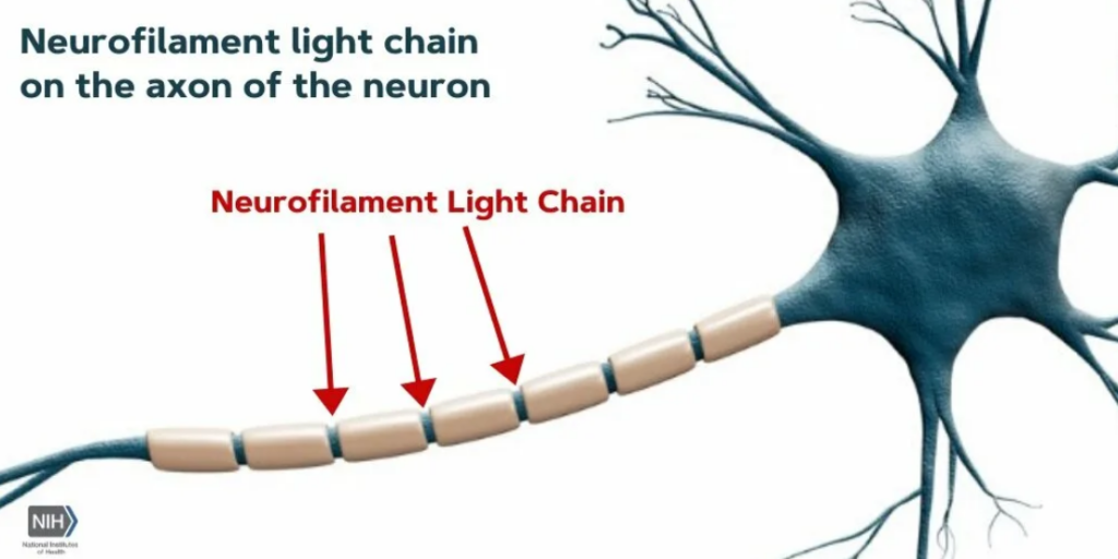
Neurofilament Light Chain (NfL) is a structural protein of neurons that is part of their cytoskeleton. It plays a key role in maintaining the shape and stability of axons (long processes of nerve cells).

Key NfL Features:

  1. Maintaining the structure of axons -provides mechanical strength and flexibility of nerve fibers.
  2. Participation in intracellular transport -helps to move substances inside the neuron.
  3. Neural tissue damage marker – when neurons are destroyed, NfL is released into the cerebrospinal fluid and blood.

When does the NfL become an important metric?

The level of NfL in the blood or cerebrospinal fluid is used as a biomarker of neurodegenerative and neurological diseases, since its increase correlates with neuronal damage.

Diseases associated with increased NfL:

  • Multiple sclerosis– MS) is a marker of demyelination activity.
  • Alzheimer ‘s disease and other dementias.
  • Parkinson ‘s disease and atypical parkinsonism.
  • Amyotrophic lateral sclerosis (ALS).
  • Traumatic brain injuries and strokes.
  • HIV-associated neurocognitive dysfunction.

Symptoms associated with Changing NfL Levels

NfL is not a cause of disease, but an indicator of neuronal damage. Therefore, ‘symptoms’ refer to the main diseases:

With increased NfL (neurodegeneration or injury):

  • Cognitive disorders (problems with memory, thinking).
  • Muscle weakness, tremor, poor coordination.
  • Numbness, tingling in the extremities (in MS).
  • Progressive dementia (with Alzheimer’s).

NfL Deficit

A physiologically low level of NfL is normal, but a complete lack of protein is impossible, since it is necessary for the structure of neurons.


NfL norm in a Blood test

NfL levels are measured in picograms per milliliter (pg / ml). The reference values depend on the age and method of analysis (for example, the Simoa method is the most sensitive).

Approximate standards:

AgeBlood NfL level (pg / ml)
< 40 years old<10
40-60 years old<15
> 60 years old<20–30

Important:

  • Children have a higher level of NfL than adults (due to the active development of the nervous system).
  • In neurological diseases, the values can exceed 50-100 pg / ml and even reach 1000 pg / ml (in late-stage ALS).

How is the analysis performed?

  1. Material: blood or CSF (cerebrospinal fluid).
  2. Method: immunoassay using antibodies (for example, Single Molecule Array-Simoa).
  3. When they are assigned:
  • Diagnosis and monitoring of multiple sclerosis.
  • Assessment of the progression of neurodegenerative diseases.
  • Monitoring the effectiveness of therapy.

What to do with an increased NfL?

  1. Consult a neurologist to find the cause (MRI, lumbar puncture, etc.).
  2. Treatment of the underlying disease (for example, immunotherapy for MS).
  3. Dynamic follow – up-repeated tests to assess the rate of progression.

Conclusion

  • NfL protein is a marker of neuronal damage, not an independent disease.
  • The norm: depends on the age, on average < 10-30 pg / ml in the blood.
  • An increase indicates neurodegeneration, trauma, or autoimmune processes (MS, ALS, Alzheimer’s).
  • NfL analysis is used in neurology for early diagnosis and disease control.

If the NfL level is elevated, an in-depth examination by a specialist is required.